How it works
From image to decision, in real time.
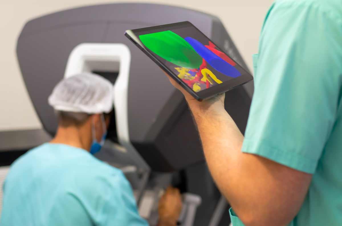
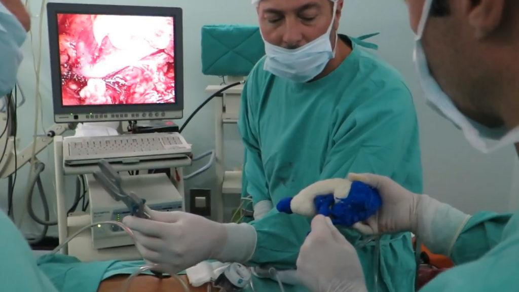
The process begins with the patient's contrast-enhanced CT. The MIRAI 3D team processes the image, segments the kidney, arteries, veins and tumor mass, and builds a volumetric biomodel. Before surgery, the biomodel is validated with the surgeon in a dedicated session.
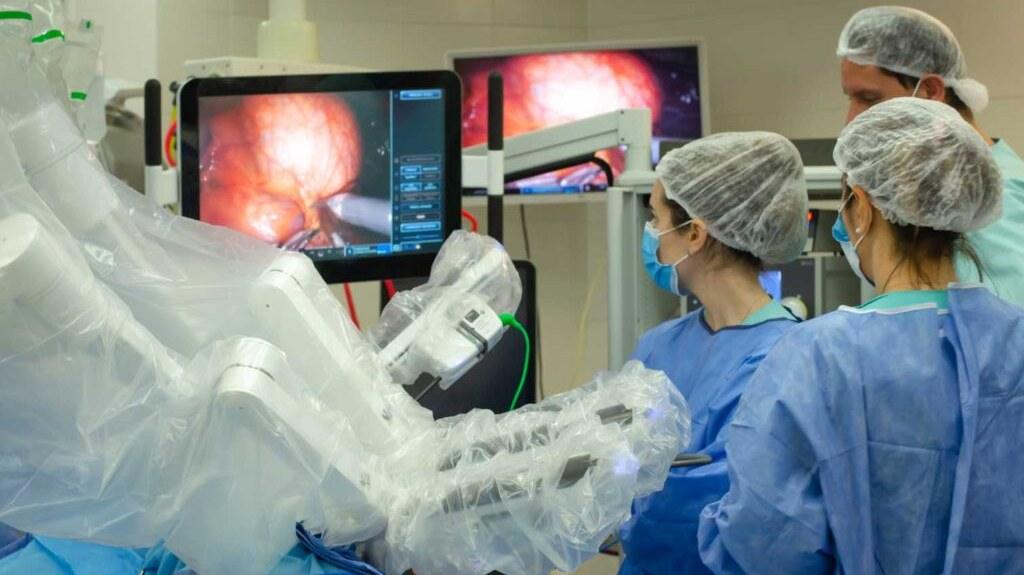
In the OR, the auxiliary screen shows the biomodel. The surgeon manipulates it with touchless gestures, which preserves the sterile field. Rotation, zoom and overlay with the robotic endoscopic image are immediate. Selective-clamping planning is confirmed or adjusted before proceeding.